Processus de recherche
9 figures issues de recherches évaluées par des pairs
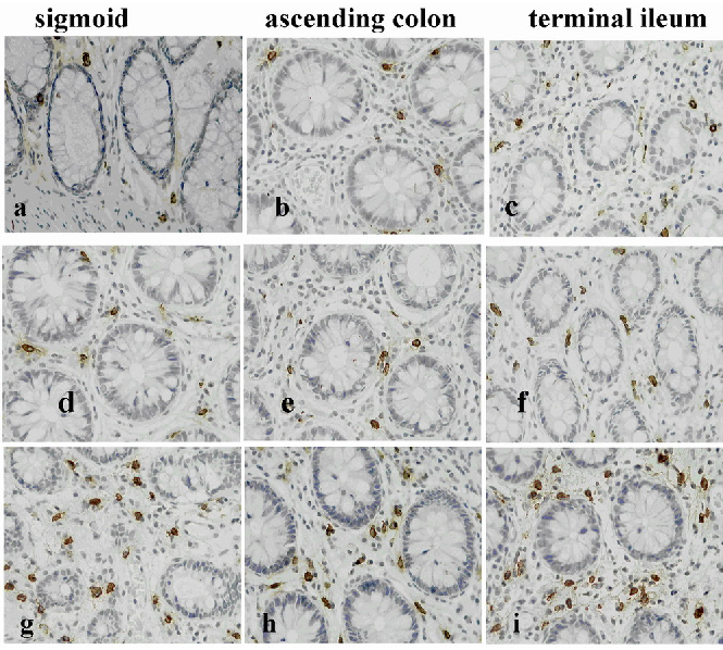
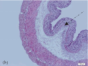
Representative photomicrographs of tryptase-positive mast cells in colonic mucosa compare healthy controls with IBS-D patients, suggesting mast cell involvement in lactose intolerance symptomatology.
Lactose Intolerance in Adults: Biological Mechanism and Dietary Management.
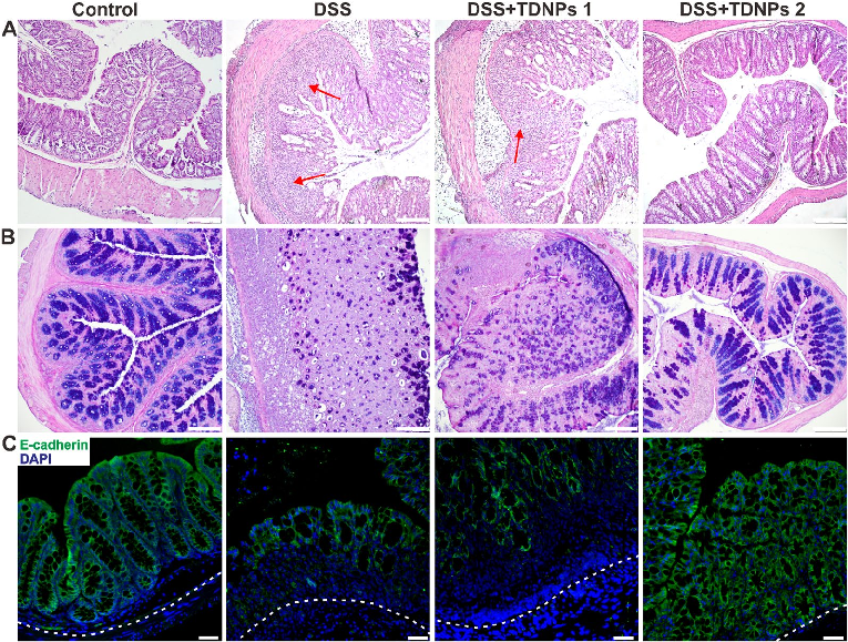
Histological examination with H&E staining reveals markedly reduced inflammatory cell infiltration and preserved goblet cell density in TDNPs 2-treated colitic mice. Colonic tissue architecture remains largely intact compared to severe disruption in untreated animals.
Oral administration of turmeric-derived exosome-like nanovesicles with anti-inflammatory and pro-resolving bioactions for …
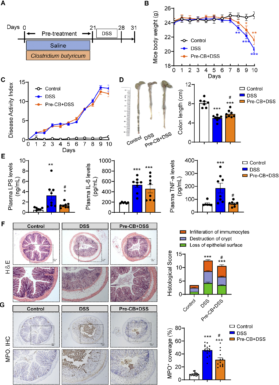
Histological analysis of colonic tissue reveals the extent of inflammatory infiltration and mucosal damage. This figure compares histopathological scores across treatment groups in the acute experimental colitis model.
Clostridium butyricum and Its Derived Extracellular Vesicles Modulate Gut Homeostasis and Ameliorate …
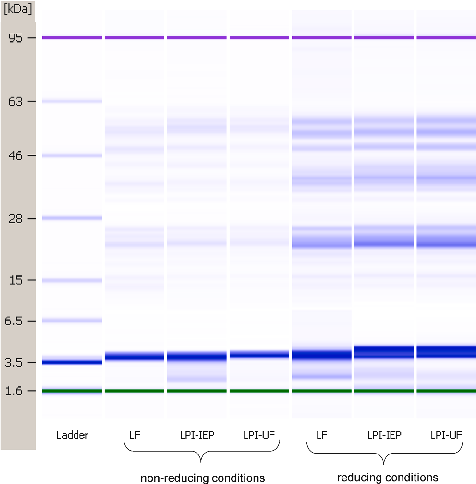
Gel-like electrophoresis images compare the protein molecular weight distribution of lentil protein sources under reducing and non-reducing conditions, revealing the impact of processing methods on protein subunit composition.
Nutritional and anti-nutritional properties of lentil (Lens culinaris) protein isolates prepared by …
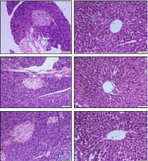
Histological examination of pancreatic and hepatic tissue compares morphological changes between vitamin A-sufficient and deficient mice.
Changes in Intestinal Microbiota Are Associated with Islet Function in a Mouse …
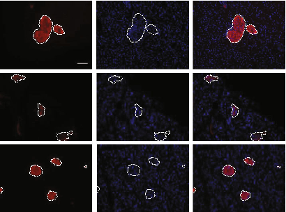
Immunofluorescence staining for insulin and DAPI in pancreatic islets reveals altered islet architecture and reduced insulin-positive area in vitamin A-deficient mice.
Changes in Intestinal Microbiota Are Associated with Islet Function in a Mouse …
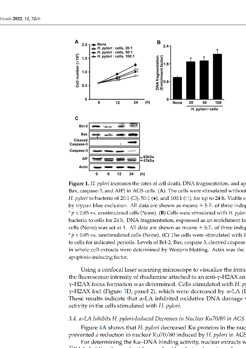
Immunofluorescence imaging of Ku70/Ku80 localization in gastric epithelial cells, showing nuclear protein retention in alpha-lipoic acid-treated cells despite H. pylori infection.
α-Lipoic Acid Inhibits Apoptosis by Suppressing the Loss of Ku Proteins in …
Liver histopathology from colitis-induced mice with and without oral NAC supplementation, evaluating extraintestinal hepatic manifestations including steatosis and inflammatory infiltration.
Extraintestinal Manifestations in Induced Colitis: Controversial Effects of N-Acetylcysteine on Colon, Liver, …
Kidney tissue histology showing renal changes associated with induced colitis, and the effects of N-acetylcysteine on renal inflammation and oxidative damage markers.
Extraintestinal Manifestations in Induced Colitis: Controversial Effects of N-Acetylcysteine on Colon, Liver, …