Proses Penelitian
211 gambar dari penelitian yang ditinjau oleh rekan sejawat
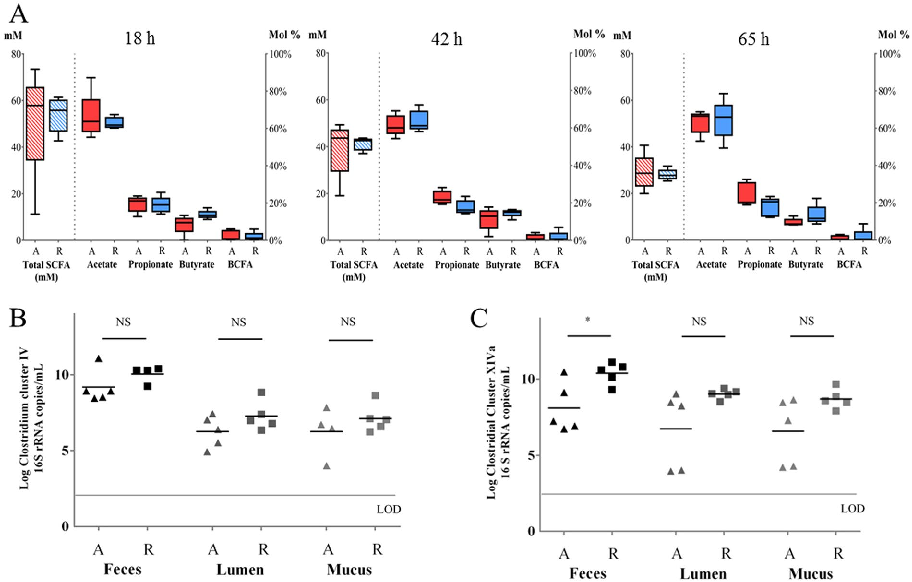
Metabolomic analysis of fecal samples following inulin intervention reveals significant changes in short-chain fatty acid profiles, particularly increased butyrate and propionate concentrations. These metabolic shifts correlate with specific microbial community changes.
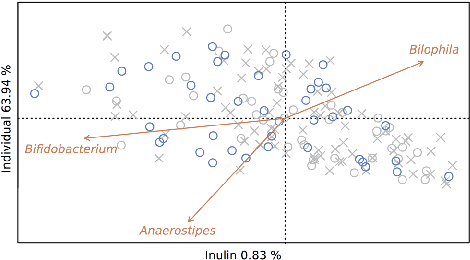
Prebiotic inulin-type fructans induce specific changes in the human gut microbiota.
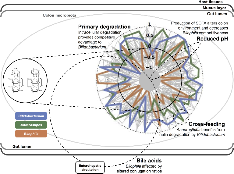
Gut microbiota composition changes induced by inulin-type fructans are characterized by increased abundance of Bifidobacterium, Faecalibacterium, and Anaerostipes, alongside decreased levels of Bilophila. These shifts are associated with altered fecal metabolite profiles.
Prebiotic inulin-type fructans induce specific changes in the human gut microbiota.
Funnel plot assessment for publication bias in the probiotic meta-analysis indicates generally symmetric distribution of effect sizes around the pooled estimate.
A meta-analysis of probiotic efficacy for gastrointestinal diseases.
Butyrate and other short-chain fatty acid concentrations measured in the in vitro fermentation model show significant increases following supplementation with targeted bacterial strains.
Butyrate-producing bacteria supplemented in vitro to Crohn's disease patient microbiota increased butyrate …

Relative abundance shifts in key bacterial taxa after supplementation are displayed, highlighting the engraftment success of introduced butyrate producers.
Butyrate-producing bacteria supplemented in vitro to Crohn's disease patient microbiota increased butyrate …
pH changes and metabolite profiles in the fermentation vessel over time demonstrate the metabolic impact of butyrate-producing bacterial supplementation.
Butyrate-producing bacteria supplemented in vitro to Crohn's disease patient microbiota increased butyrate …
Diversity indices comparing baseline Crohn's disease microbiota to supplemented samples suggest partial restoration of microbial community richness.
Butyrate-producing bacteria supplemented in vitro to Crohn's disease patient microbiota increased butyrate …
Correlation analysis between butyrate concentration increases and shifts in specific bacterial genera reveals ecological relationships within the supplemented microbiota.
Butyrate-producing bacteria supplemented in vitro to Crohn's disease patient microbiota increased butyrate …
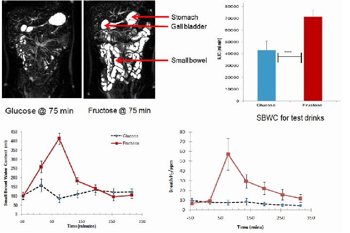
Small bowel water content and breath hydrogen concentrations after consuming glucose and fructose drinks are plotted over time, providing reference data for comparison with lactose challenge tests.
Lactose Intolerance in Adults: Biological Mechanism and Dietary Management.
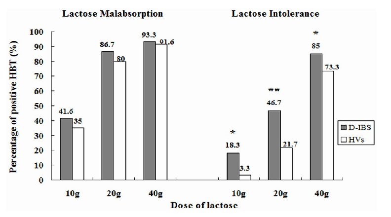
Prevalence of lactose malabsorption versus symptomatic lactose intolerance in IBS-D patients is compared, demonstrating that malabsorption does not invariably produce clinical symptoms.
Lactose Intolerance in Adults: Biological Mechanism and Dietary Management.
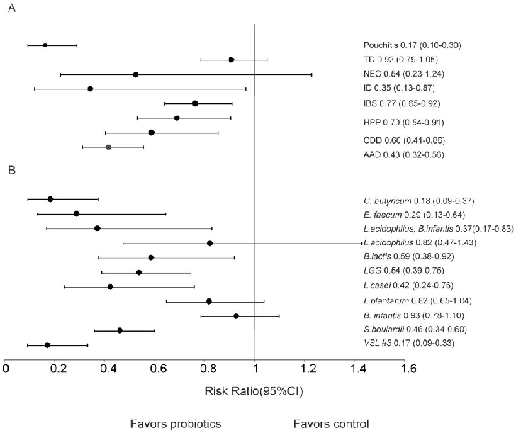
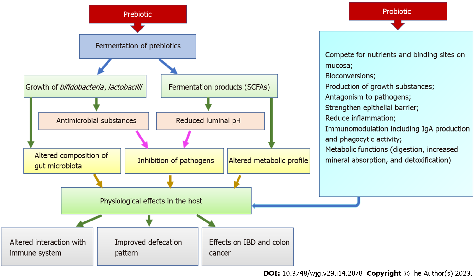
Clinical evidence for probiotic efficacy in ulcerative colitis is stronger than for Crohn's disease, with certain multi-strain formulations showing benefit in maintaining remission. Strain-specific effects are critical, as not all probiotics demonstrate equivalent therapeutic potential.
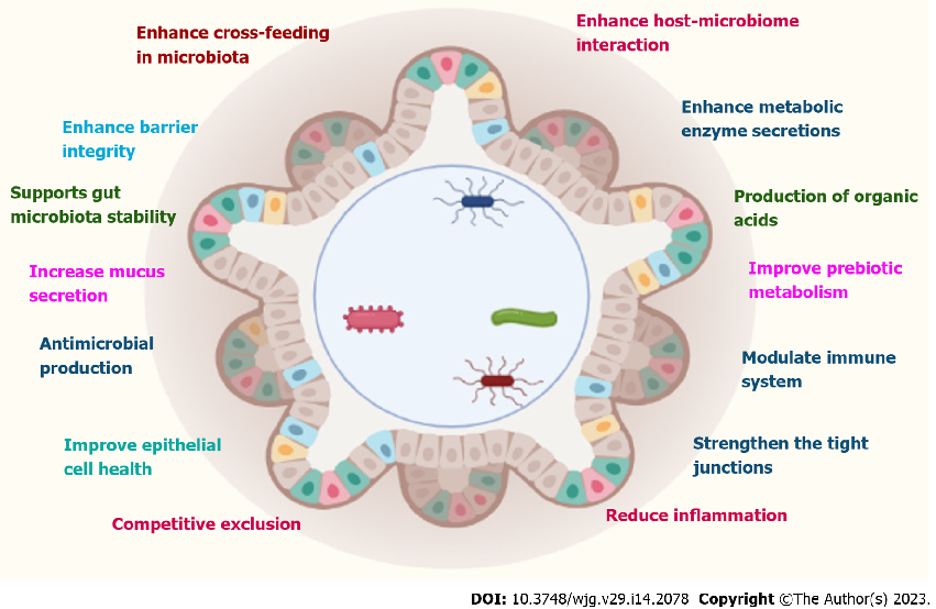
Role of prebiotics, probiotics, and synbiotics in management of inflammatory bowel disease: …
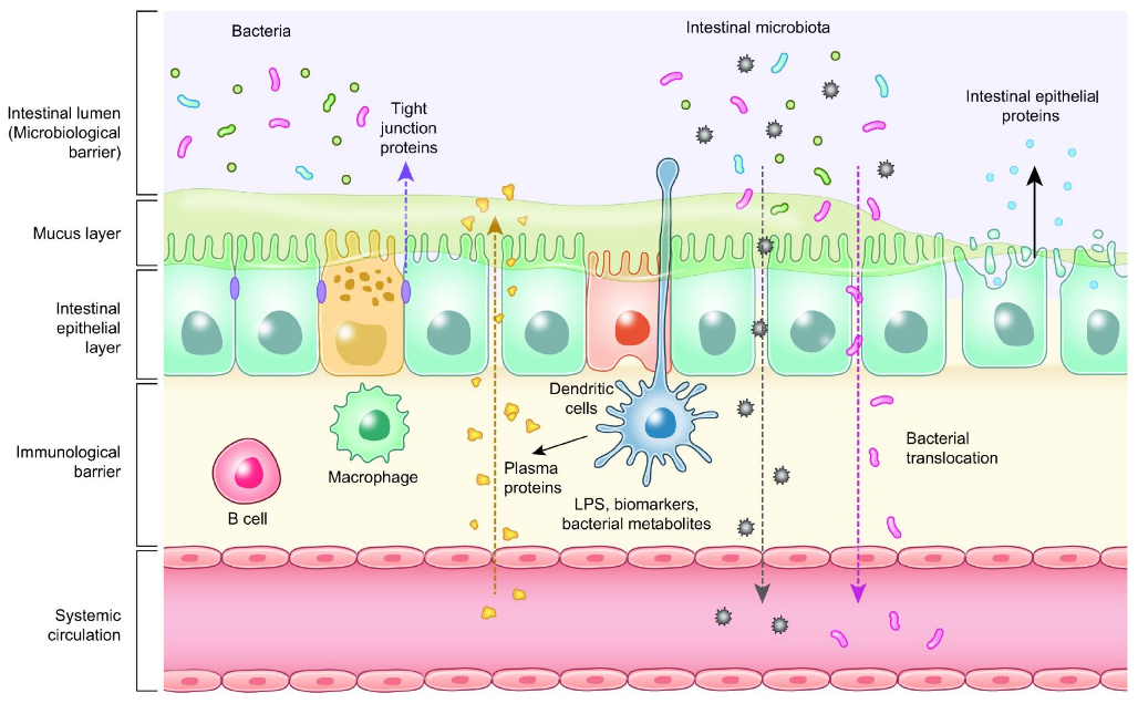
Gut microbiota composition in IBD patients is characterized by reduced diversity, decreased Firmicutes abundance, and increased representation of potentially pathogenic Proteobacteria. Probiotic interventions aim to partially normalize these patterns.
Role of prebiotics, probiotics, and synbiotics in management of inflammatory bowel disease: …

Fecal calprotectin and other inflammatory biomarkers may serve as objective measures for monitoring probiotic treatment response in IBD patients. Reductions in these markers correlate with clinical improvement in several studies.
Role of prebiotics, probiotics, and synbiotics in management of inflammatory bowel disease: …
Essential oil compounds from oregano, thyme, and cinnamon demonstrate dose-dependent antimicrobial activity against common poultry pathogens. Their mode of action involves disrupting bacterial cell membrane integrity.
Probiotics, Prebiotics, and Phytogenic Substances for Optimizing Gut Health in Poultry.
Synbiotic combinations in poultry nutrition show additive or synergistic effects compared to individual probiotic or prebiotic supplementation. Performance metrics including feed conversion ratio and weight gain may improve with optimized formulations.
Probiotics, Prebiotics, and Phytogenic Substances for Optimizing Gut Health in Poultry.
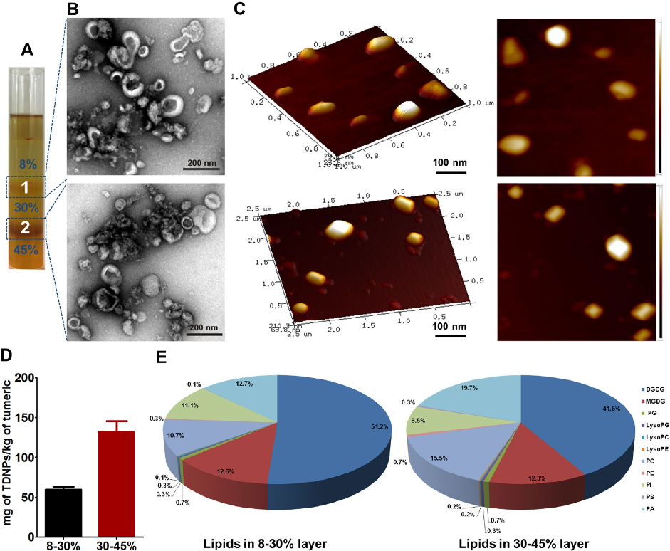
Characterization of turmeric-derived nanoparticles reveals two distinct bands (TDNPs 1 and TDNPs 2) at the 8%/30% and 30%/45% sucrose gradient interfaces, respectively. TDNPs 2 demonstrate appropriate size distribution and surface charge for oral drug delivery applications.
Oral administration of turmeric-derived exosome-like nanovesicles with anti-inflammatory and pro-resolving bioactions for …
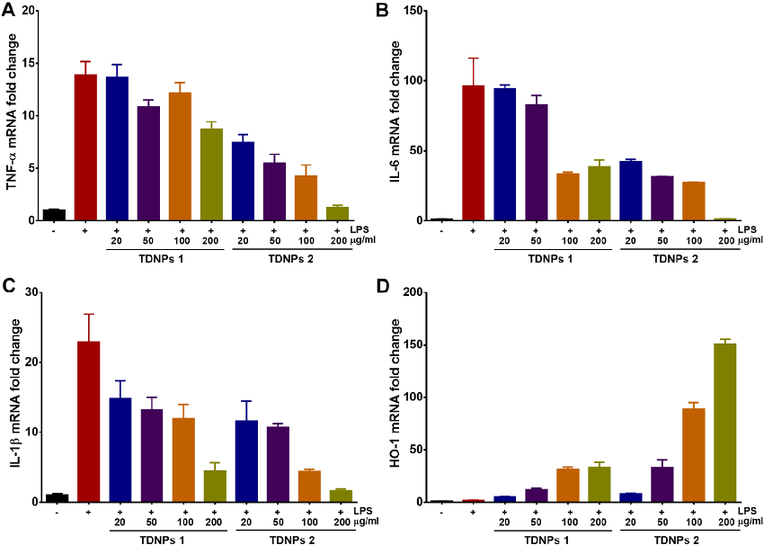
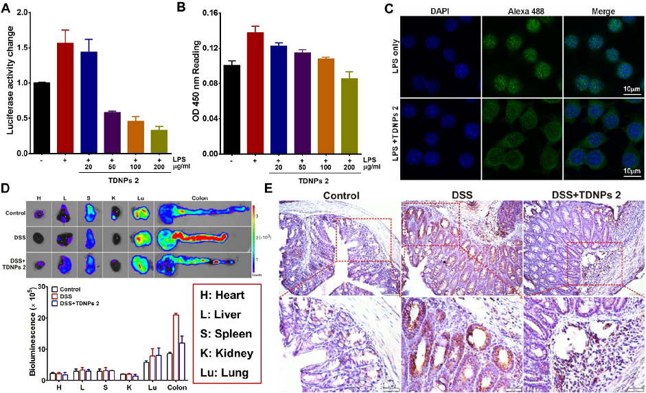
In vitro assessment of turmeric-derived nanovesicles demonstrates anti-inflammatory activity, including suppression of pro-inflammatory cytokine production in activated macrophages. Dose-dependent reductions in TNF-alpha and IL-6 secretion are observed.
Oral administration of turmeric-derived exosome-like nanovesicles with anti-inflammatory and pro-resolving bioactions for …
Oral TDNPs 2 administration significantly attenuates disease activity index scores and colon shortening in DSS-induced colitis mice. Body weight recovery is also improved compared to untreated colitis controls.
Oral administration of turmeric-derived exosome-like nanovesicles with anti-inflammatory and pro-resolving bioactions for …
Intestinal permeability assessment indicates that TDNPs 2 treatment preserves gut barrier integrity in colitis mice. Tight junction protein expression, including ZO-1 and occludin, is maintained at near-normal levels.
Oral administration of turmeric-derived exosome-like nanovesicles with anti-inflammatory and pro-resolving bioactions for …
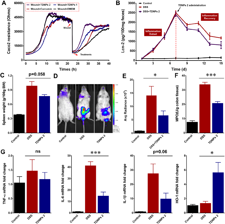
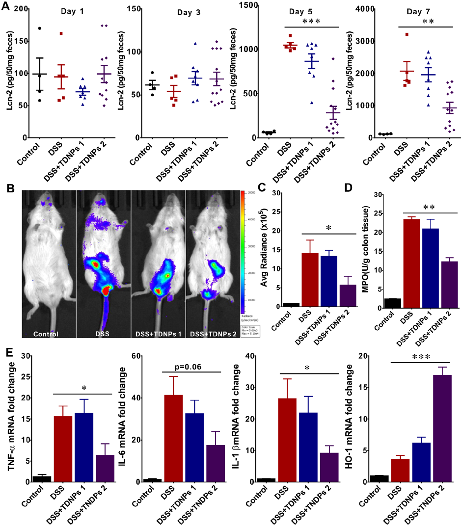
Wound healing assays and fecal lipocalin-2 quantification demonstrate that TDNPs 2 accelerate resolution of intestinal inflammation. Lcn-2 levels, a sensitive marker of intestinal inflammation, decrease significantly with treatment.
Oral administration of turmeric-derived exosome-like nanovesicles with anti-inflammatory and pro-resolving bioactions for …
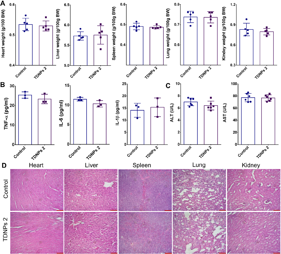
Biocompatibility evaluation shows no significant changes in vital organ weights, pro-inflammatory cytokines, or liver function indicators in TDNPs 2-treated mice. H&E staining of major organs confirms absence of toxicity.
Oral administration of turmeric-derived exosome-like nanovesicles with anti-inflammatory and pro-resolving bioactions for …
TDNPs 2 exert their protective effect at least partly through inactivation of the NF-kB signaling pathway. Reduced phospho-NF-kB p65 expression and decreased nuclear translocation indicate suppression of this key inflammatory cascade.
Oral administration of turmeric-derived exosome-like nanovesicles with anti-inflammatory and pro-resolving bioactions for …
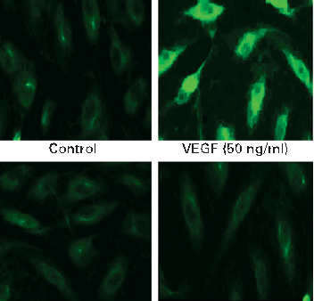
COX-2 protein expression is upregulated by VEGF stimulation in intestinal endothelial cells, and curcumin effectively blocks this induction. Prostaglandin E2 production follows a similar pattern of inhibition.
Curcumin inhibits VEGF-mediated angiogenesis in human intestinal microvascular endothelial cells through COX-2 …
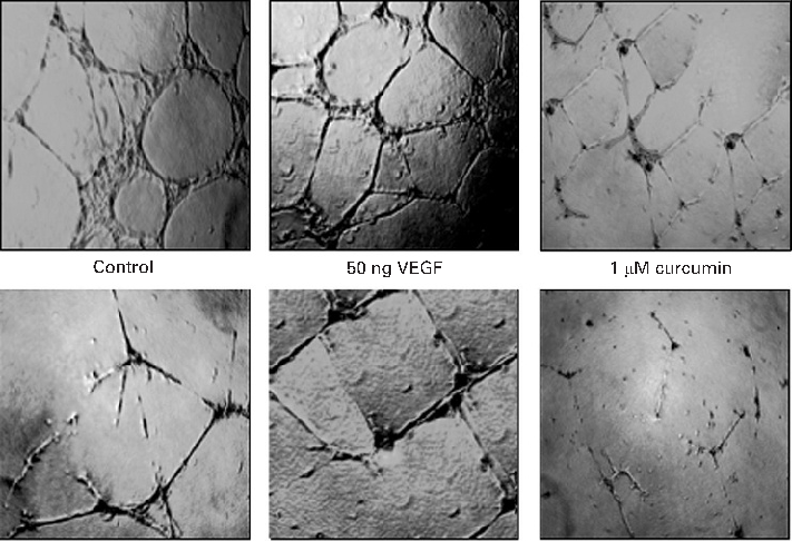
MAPK signaling pathway activation by VEGF is attenuated by curcumin in a time- and dose-dependent manner. Phosphorylation of ERK, p38, and JNK is markedly reduced in curcumin-treated endothelial cells.
Curcumin inhibits VEGF-mediated angiogenesis in human intestinal microvascular endothelial cells through COX-2 …
Halaman 1 dari 9