Descripción
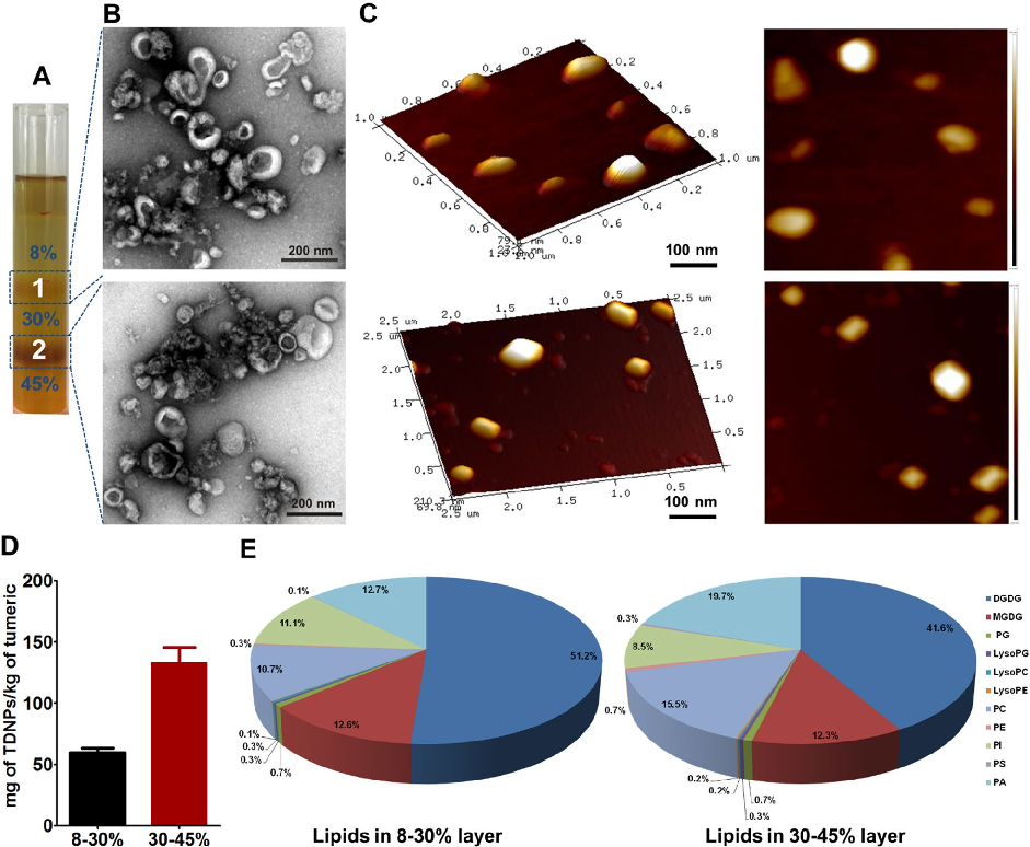
Characterization of turmeric-derived nanoparticles reveals two distinct bands (TDNPs 1 and TDNPs 2) at the 8%/30% and 30%/45% sucrose gradient interfaces, respectively. TDNPs 2 demonstrate appropriate size distribution and surface charge for oral drug delivery applications.
More Figures from This Paper
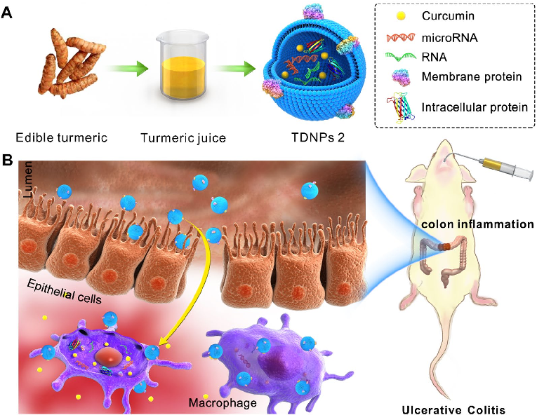
Figure 1
Turmeric-derived nanoparticles (TDNPs 2) are isolated through sucrose gradient ultracentrifugation and administered orally to target inflamed colonic tissue in a murine colitis model. The schematic outlines the isolation workflow from edible turmeric to purified nanovesicles.
diagram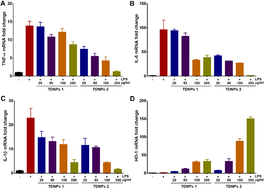
Figure 3
In vitro assessment of turmeric-derived nanovesicles demonstrates anti-inflammatory activity, including suppression of pro-inflammatory cytokine production in activated macrophages. Dose-dependent reductions in TNF-alpha and IL-6 secretion are observed.
chart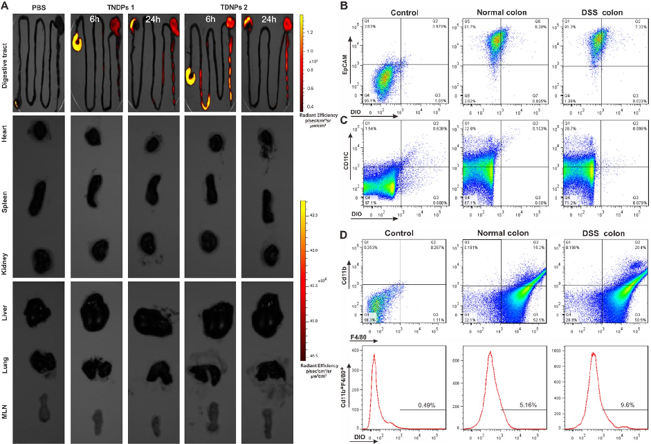
Figure 4
Biodistribution imaging using IVIS Spectrum reveals that TDNPs 2 preferentially accumulate in inflamed colonic tissue following oral administration. Fluorescence signals are minimal in non-target organs including heart, liver, spleen, lung, and kidney.
photograph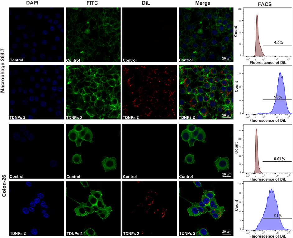
Figure 5
Oral TDNPs 2 administration significantly attenuates disease activity index scores and colon shortening in DSS-induced colitis mice. Body weight recovery is also improved compared to untreated colitis controls.
chart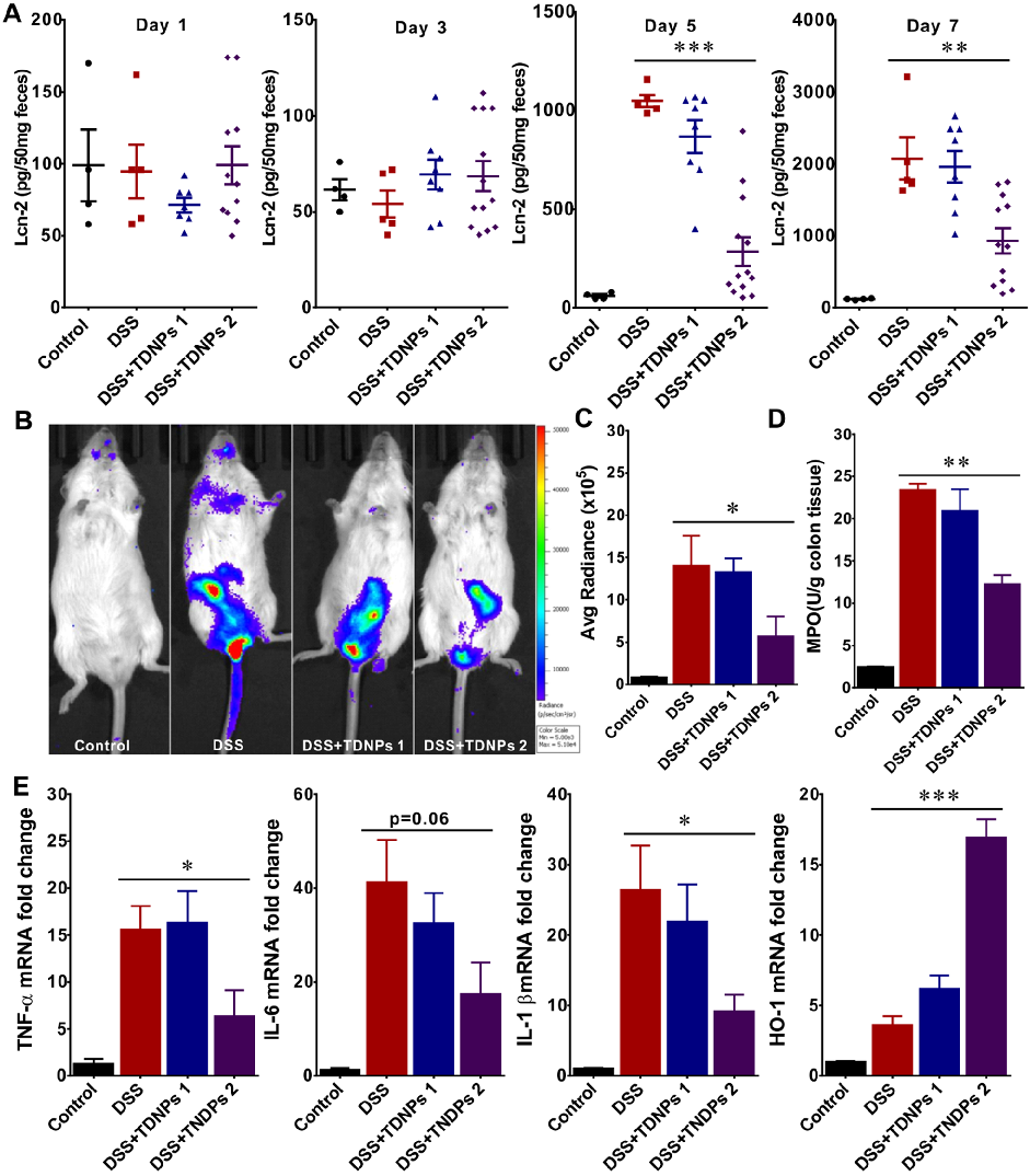
Figure 6
Intestinal permeability assessment indicates that TDNPs 2 treatment preserves gut barrier integrity in colitis mice. Tight junction protein expression, including ZO-1 and occludin, is maintained at near-normal levels.
chart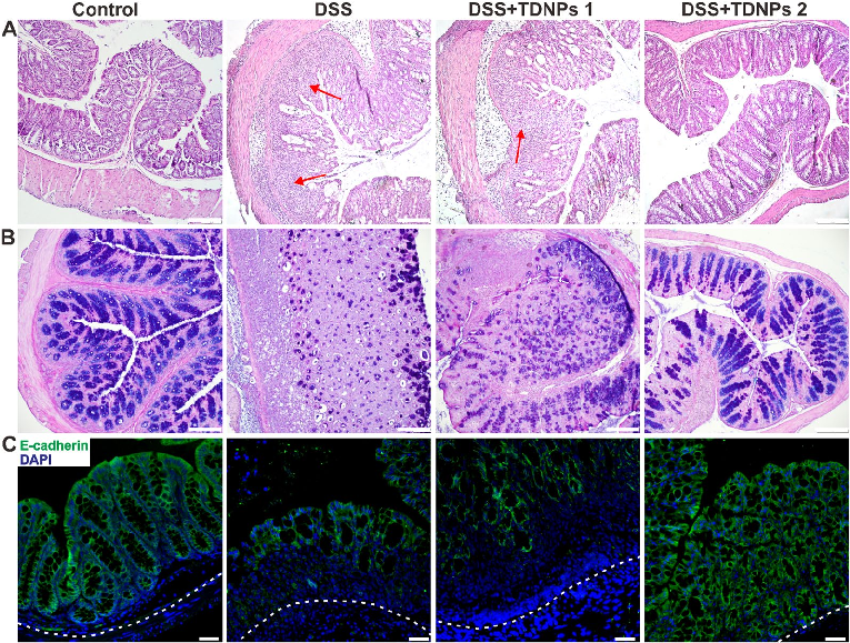
Figure 7
Histological examination with H&E staining reveals markedly reduced inflammatory cell infiltration and preserved goblet cell density in TDNPs 2-treated colitic mice. Colonic tissue architecture remains largely intact compared to severe disruption in untreated animals.
micrographFigure 2
ChartSource Paper
Oral administration of turmeric-derived exosome-like nanovesicles with anti-inflammatory and pro-resolving bioactions for murine colitis therapy.Cite This Figure
 > Source: Cui Liu et al. "Oral administration of turmeric-derived exosome-like nanovesicles with anti-infl." *Journal of nanobiotechnology*, 2022. PMID: [35488343](https://pubmed.ncbi.nlm.nih.gov/35488343/)
<figure>
  <img src="https://pdfs.citedhealth.com/figures/35488343/89.png" alt="Characterization of turmeric-derived nanoparticles reveals two distinct bands (TDNPs 1 and TDNPs 2) at the 8%/30% and 30%/45% sucrose gradient interfaces, respectively. TDNPs 2 demonstrate appropriate size distribution and surface charge for oral drug delivery applications." />
  <figcaption>Figure 2. Characterization of turmeric-derived nanoparticles reveals two distinct bands (TDNPs 1 and TDNPs 2) at the 8%/30% and 30%/45% sucrose gradient interfaces, respectively. TDNPs 2 demonstrate appropriate size distribution and surface charge for oral drug delivery applications.<br>  Source: Cui Liu et al. "Oral administration of turmeric-derived exosome-like nanovesicles with anti-infl." <em>Journal of nanobiotechnology</em>, 2022. PMID: <a href="https://pubmed.ncbi.nlm.nih.gov/35488343/">35488343</a></figcaption>
</figure>